University of South Florida
Cohort 2012 SRNA
Senior Project
EST. 2014
"By failing to prepare, you are preparing to fail"

Flexible Fiberoptic Bronchoscope
Indications
-
Used in a variety of clinical scenerios when difficult intubation is anticipated such as lower and/or upper airway obstruction, unstable cervical spine injuries, risk of dental injury, and awake intubations
-
Utilized to identify structures below the level of the vocal cords
-
Can be performed before inducing general anesthesia eliminating the risk of failed intubation or failed ventilation in patients who have been anesthetized
Contraindications
-
Hypoxia
-
Copious airway secretions unrelieved with suction or antisialagogues
-
Bleeding from the airway unrelieved with suction
-
Local anesthetic allergy if performing awake
-
Inability to cooperate if performing awake
Technology
-
Constructed of coded glass fibers that transmit light and images by internal reflection
-
Insertion shaft contains two bundles of fibers
-
each contains 10,000 to 15,000 fibers
-
one bundle transmits light from the light source
-
the other bundle provides a high resolution image
-
-
The shaft is directionally manipulated with an angulation wire
-
Aspiration channels allow for suctioning of secretions, insufflation of oxygen, or injection of fluids
Technique
-
Fully explain the procedure and ensure patient cooperation
-
Can be performed nasally or orally while awake or asleep
-
Nasal - the nasal mucous should be anesthetized and vasoconstricted with lidocaine and phenylephrine
-
Oral - topical local anesthetics or nerve blocks may be performed
-
-
An antisialagogues can be used to decrease secretions
-
Pre-procedure sedation may be utilized if appropriate
-
Place an endotracheal tube over the shaft of the bronchoscope
-
Insert the bronchoscope into the airway
-
Advance and manipulate bronchoscope until larynx and vocal cords are in view
-
Continue to advance bronchoscope past the vocal cords until visualization of carina
-
While firmly holding bronchoscope with carina in view, slide endotracheal tube off bronchoscope
-
Once the endotracheal tube is properly placed above the carina, inflate cuff and withdraw bronchoscope
Tips and Trick for a Successful Intubation
-
Numerous maneuvers may help open the airway and enlarge the channel for the fiberscope.
-
These include a jaw thrust, pulling on the tongue with a gauze pad, and inserting a laryngoscope to distract the tongue and jaw.
-
-
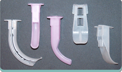
Specialized fiberoptic oral airways (i.e., Berman, Ovassapian, and Williams) are very effective for getting around the base of the tongue, protecting the scope from biting, and creating an open channel for the scope to pass through.
-
If resistance is met when advancing the endotracheal tube, rotate it 90° counterclockwise while continuing to advance


